
The human heart is one of the most important organs present in the body.
Heart – is a small hollow muscular organ , shaped somewhat like an upside-down pear , about the size of a fist, that is responsible for pumping blood throughout the blood vessels to various parts of the body, called the cardiovascular system.
The weight of the human heart can be anything in between 250 -400 grams. Heart is found superior to diaphragm, posterior to sternum, slightly left or center of the chest and between the lungs and is specifically located in the mediastinum of thoracic cavity. The average human heart beats at 72 bpm and pumps approximately 4.7-5.7 litres of blood per minute and more if you are running.
FOUR CHAMBERS OF THE HEART AND THEIR FUNCTIONS:
* two atria ( upper chambers) and
* two ventricles(lower chambers)
- The right atrium receives deoxygenated blood from the veins and pumps it to the right ventricle.
- The right ventricle receives deoxygenated blood from the right atrium and pumps it to the lungs, where it is oxygenation takes place..
- The left atrium receives oxygenated blood from the lungs and pumps it to the left ventricle.
- The left ventricle (the strongest chamber) receives oxygenated blood from the left atrium and pumps oxygen-rich blood to the rest of the body via aorta.
LAYERS OF THE HEART AND THEIR FUNCTIONS:
- Pericardium
The pericardium is the double walled , fluid filled sac that contains the heart and the proximal ends of the aorta, vena cava, and the pulmonary artery.
FUNCTIONS:
Holds the heart in the chest cavity.
Prevents the heart from becoming too large when blood volume increases.
Limits heart movements. - Myocardium
The myocardium is the thickest and middle layer of the heart. It is made of cardiac muscles, responsible for the heart’s pumping of blood to the entire body. The coronary arteries supply blood and oxygen to myocardium. - Endocardium
The endocardium is the thin inner layer of the heart. Its main function is to allow the smooth flow of blood through the heart.
FOUR VALVES OF THE HEART AND THEIR FUNCTIONS:
Function of the valves:
Controls the direction of blood flow,thus preventing the backward flow of blood.
Blood passes through a valve before leaving the four chamber of the heart that collect and pump blood. Valves are made of flaps, or leaflets that act as one-way inlets for blood coming into a ventricle and one-way outlets for blood leaving a ventricle. The four heart valves include the following:
- Tricuspid valve -( atrioventricular valve) located between the right atrium and the right ventricle( right side of the heart). The tricuspid valve is the first valve where blood flows through in the heart. It is made of three flaps, or leaflets, that work together to stop and start the flow of blood.
- Pulmonary valve – is the second valve of the heart referred as semilunar valve, because of its shape. It is located between the right ventricle and the pulmonary artery, which carries blood to the lungs. When the right ventricle contracts, the tricuspid valve opens, allowing blood to flow to the lungs.
- Mitral valve or bicuspid valve (atrioventricular valve) is the third valve of the heart. located between the left atrium and the left ventricle. The mitral valve is composed of two flaps, or leaflets, that prevent blood from flowing into the ventricle prematurely. When the atrium contracts, the mitral valve opens, allowing blood to move into the ventricle.
- Aortic valve -The aortic valve is the fourth heart valve, located between the left ventricle and the aorta . The valve is composed of three flaps or leaflets, working together to prevent blood from entering the aorta prematurely. The aortic valve opens when the ventricle contracts, allowing blood to move from the heart to the rest of the body.
HEART ABNORMALITIES:
Coronary Artery Disease (CAD) –is the most significant threat to the myocardium, as occlusions in the coronary arteries deprive the myocardium of blood and oxygen. Coronary artery disease (CAD) also known as atherosclerotic heart disease, coronary heart disease,or ischemic heart disease (IHD), is the most common type of heart disease and cause of heart attacks.
Stable Angina pectoris – is chest pain, that commonly occur during activity or stress, due to obstruction or spasm of the coronary arteries. The symptoms are predictable. Narrowed or occluded coronary arteries caused by atherosclerosis or by a blood clot, prevent the heart from receiving the right amout of oxygen needed for strenous activity thus, causing chest pain or chest discomfort with exertion. Typically, the symptoms get better with rest. Stable angina is not as serious as unstable angina, but it can be very painful or uncomfortable.
Unstable Angina, also called acute coronary insufficiency , symptoms are unpredictable and do not correlate to increased demands on the heart such as physical activity may place. It typically occurs at rest and has a sudden onset, sudden worsening, and stuttering recurrence over days and weeks. This is an emergency situation as it can precede a heart attack, serious abnormal heart rhythm, or cardiac arrest. Angioplasty or CABG is often the most viable treatment options.
Management reference: Wikipedia
Nitroglycerin can be used immediately to widen the coronary arteries and help increase blood flow to the heart. In addition, nitroglycerin causes peripheral venous and artery dilation reducing cardiac preload and afterload. These reductions allow for decrease myocardial oxygen demand. Antiplatelet drugs such as aspirin and clopidogrel can help reduce the progression of plaque formation, as well as combining these with an anticoagulant such as a low molecular weight heparin.
Myocardial Infarction (heart attack)- destruction of heart tissue resulting from insufficient blood supply to the heart muscle. It happens when one of the coronary arteries that supplies blood to the heart develops a blockage or occlusion.
Symptoms:
Sudden pain, pressure, fullness, squeezing in the center of the chest , and may radiates to the left shoulder, to the left side of the neck, to the left arm and sometimes to the jaws. Patient may have shortness of breath, sudden weakness, dizziness, diaphoresis, nausea, vomiting, and anxiety.
Risk Factors:
History of cardiovascular disease, old age, tobacco smoking, high cholesterol, diabetes, high blood pressure, lack of physical activity, obesity, chronic kidney disease , excessive alcohol intake, and chronic high stress levels.
Tests:
Electrocardiogram(ECGs). Blood test for( Cardiac Enzymes) creatinine kinase (CK-MB) and troponin.
Arrhythmia (dysrhythmia): An irregular heartbeat due to changes in the conduction of electrical impulses through the heart. Some arrhythmias are benign, some are life-threatening.
Symptoms:
- Chest pain
- Fainting
- Change in level of consciousness or lethargy
- Lightheadedness or dizziness
- Pale skin
- Rapid breathing (tachypnea)
- Short of Breath
- Rapid heart rate (tachycardia)
- Skipping beats (changes in the pattern of the pulse)
- Slow heartbeat (bradycardia)
- Sweating
Some common causes of abnormal heartbeats are:
- Abnormal levels of potassium or other substances
- Heart attack, or a damaged heart muscle from a past heart attack
- Congenital Heart disease
- Heart failure or an enlarged heart
- Hyperthyroidism or Overactive thyroid gland
- Alcohol, caffeine, or stimulants such as amphetamines
- Beta-blockers
- Cigarette smoking (nicotine)
- Medicines used for depression or psychosis
- Some prescription drugs
Congestive Heart Failure(CHF) is a condition in which the heart is either too weak or too stiff to effectively pump out enough oxygen rich blood through the body.
Common symptoms:
- Shortness of breath
- Leg swelling
Cardiomyopathy : A disease of the heart muscle (myocardium) in which the heart is abnormally enlarged, thickened, or rigid. Thus, the heart’s ability to pump blood is weakened.
Common symptoms:
- Shortness of breath
- Chest pain
- Fatigue
Myocarditis: Inflammation of the heart muscle (myocardium), most often due to a viral infection.
Symptoms may include one or more of these:
- Shortness of breath
- Abnormal heartbeat
- Light-headedness
- Chest pain
- Fatigue
- Fever
- Muscle aches,
- Sore Throat
- Headache
- Diarrhea
- Painful joints
- Swollen joints, legs, or neck veins
Pericarditis: Inflammation of the fibrous sac surrounding the heart (pericardium). Viral infections, kidney failure, and autoimmune conditions are common causes.
Symptoms:
- Chest pain
- Dry cough
- Fever
- Fatigue
- Anxiety
Pericardial Effusion: Abnormal accumulation of fluid between the lining of the heart (pericardium) and the heart itself. Often, this is due to pericarditis.
Symptoms of Pericardial Effusion related to Pericarditis:
- Fever
- Chest pain
- Fatigue
- Muscle aches
- Shortness of breath
- Nausea, vomiting, and diarrhea
Serious pericardial effusions may cause symptoms including:
- Shortness of breath
- Palpitations
- Light-headedness or fainting
- Cool, clammy skin
Atrial Fibrillation: is one of the most common arrhythmias. Atrial fibrillation is an abnormal rhythm of the heart, which is caused by abnormal electrical impulses within the atria. Atrial fibrillation reduces the ability of the atria to pump blood into the ventricles and usually causes fast heartbeat , irregular heartbeat (arrhythmia) that can lead to blood clots, stroke, heart failure and other heart-related complications
Symptoms of atrial fibrillation include (some people may have no symptoms).
- Palpitations
- Dizziness
- Fainting
- Weakness
- Fatigue
- Shortness of Breath
- Chest Pain
Pulmonary Embolism(PE): is a blockage of a major blood vessel (artery) in the lung or one of it’s branches, usually by a blood clot that has travelled from other parts of the body through bloodstream
Causes of Pulmonary Embolism
- Immobilization: Common to bed ridden pts.
- Heart Disease (such as arrhythmias)
- Travel: Prolonged sitting, such as in an airplane or a long car trip, allows the blood to sit in the legs and increases the risk of clot formation.
- Obesity
- Trauma or injury (especially to the legs)
- Recent surgery .(It is also associated with immobility and sometimes vessel damage during surgery)
- Previous history of blood clot in the legs (DVTs) or pulmonary embolism
Endocarditis: Inflammation of the inner layer of the heart, the endocardium. Usually, endocarditis is due to a serious infection of one of the heart valves.
Mitral Valve Prolapse: Normally, the mitral valve helps blood on the left side of the heart flow in one direction. It closes to keep blood from flowing backwards when the heart contracts. But in a person with a mitral valve prolapse, the valve does not close completely, and can cause blood to leak backwards from the left ventricle, through the mitral valve, and into the left atrium, when the left ventricle contracts, This is called mitral regurgitation.
BLOOD FLOW INSIDE THE HEART
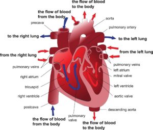
Right Side of The Heart: DEOXYGENATED BLOOD
BODY—->>Blood enters the heart via —>>, the inferior and superior vena cava ( emptying deoxygenated blood from the body)——>> into the right atrium.
As the atrium contracts, blood flows from the right atrium —->>via tricuspid valve ->>to right ventricle .
When the ventricle is full — the tricuspid valves closes.– to prevent blood from flowing backward into the right atrium —-while the ventricles contract.
As the ventricle contracts, blood leaves the heart via the pulmonic valve—>> into the pulmonary artery—>> to the lungs, where oxygenation takes place —>>The oxygenated blood returns to the heart through the pulmonary veins —->>
Left Side of the Heart: OXYGENATED BLOOD
The pulmonary vein empties oxygenated blood –from the lungs —–>>into the left atrium.
As the atrium contracts, blood flows from the left atrium —->> via Mitra Valve –>>into the left ventricle ‘
When the ventricle is full — the mitral valve closes– to prevent blood from flowing backward into the left atrium while the ventricles contracts.
As the ventricle contracts, oxygen-rich blood leaves the heart —>>via the aortic valve–—–>> aorta —>> to the BODY.
